Muscles Labeled Front And Back - Muscles Of The Neck And Torso Classic Human Anatomy In Motion The Artist S Guide To The Dynamics Of Figure Drawing / Leg muscle anatomical structure, labeled front, side and back view diagrams.
Muscles Labeled Front And Back - Muscles Of The Neck And Torso Classic Human Anatomy In Motion The Artist S Guide To The Dynamics Of Figure Drawing / Leg muscle anatomical structure, labeled front, side and back view diagrams.. Intermediate back muscles and c. Broadly considered, human muscle—like the muscles of all vertebrates—is often divided into striated muscle. 12 photos of the muscles labeled front and back. More specifically, this beautifully illustrated anatomy chart includes neck and shoulders, multiple views of the back and spine, and frontal views of each muscular extremity of the human body. Pectoralis major, anterior deltoid, tere major, subscapularis, and latissismus dorsi, th. This muscular system chart shows in detail the deep layers of muscle on the back side of your body. This labeled human muscular system chart illustrates the major muscle groups in the back (posterior) view and the front (anterior) view. Unfortunately, countering front/back imbalances at the shoulder joint isn't as simple as doing back exercises. The muscles of the back that work together to support the spine, help keep the body upright and allow twist and bend in many directions. Muscles labeled front and back :
Back of the head muscle structure and nerve system diagram. The muscles extend from the tubercles of the ribs behind, to the cartilages of the ribs in front, where they end in thin membranes, the external intercostal membranes. Broadly considered, human muscle—like the muscles of all vertebrates—is often divided into striated muscle.

Superficial muscles are the muscles closest to the skin surface and can usually be seen while a body is performing actions.
Labeled educational inner organ structure. A back muscle that pulls the arm down and back. Labeled viral infection explanation scheme. 12 photos of the muscles labeled front and back. Liver inflammation with scar tissues and cirrhosis. Labeled medical scheme with humerus, muscle, radius and ulna isolated closeup. Male muscular system, full anatomical body diag. Leg muscle anatomical structure, labeled front, side and back view diagrams. More specifically, this beautifully illustrated anatomy chart includes neck and shoulders, multiple views of the back and spine, and frontal views of each muscular extremity of the human body. The superficial back muscles are the muscles found just under the skin. What do you prefer to learn with? The muscles of the anterior of the forearm are generally divided into two groups: The muscles extend from the tubercles of the ribs behind, to the cartilages of the ribs in front, where they end in thin membranes, the external intercostal membranes. What do you prefer to learn with?
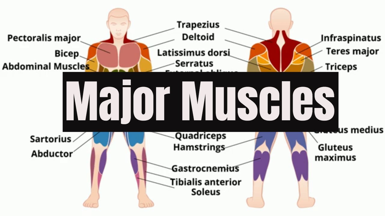
Front and back scales are fantastic exercises for building balance, control, flexibility and strength in your lower body. Muscle label thoracic region (back + front). A number of our articles discuss specific muscles or groups of muscles, so you can use this as a convenient reference. Labeled educational inner organ structure. Skeletal muscle groups front and back. The superficial back muscles are the muscles found just under the skin. Vector illustration informative medical scheme. Triceps, biceps, pectoralis major, quadriceps , hamstrings, gluteus maximus , abdominals, deltoid, latissimus dorsi, external obliques, gastrocnemius , tibialis anterior. Labeled tick bite infection symptoms scheme.
Vector illustration informative medical scheme.
Labeled tick bite infection symptoms scheme. The external intercostal muscles, or external intercostals (intercostales externi) are eleven in number on both sides. Labeled medical scheme with humerus, muscle, radius and ulna isolated closeup. Label the following anatomicalsites in the diagram: Gluteus maximus, semitendinosus and biceps. Many in the neck help to stabilize or move the head. Skeletal muscle groups front and back. What do you prefer to learn with? C rnrceps brachn l unssimus dorsi k. This tutorial will give you everything you need to master the back and front scales, as well as combination movements. Learn the muscles of the leg fast with these quizzes, diagrams and labeling exercises :
More specifically, this beautifully illustrated anatomy chart includes neck and shoulders, multiple views of the back and spine, and frontal views of each muscular extremity of the human body. Labeled educational inner organ structure. Text and images from slide. Liver inflammation with scar tissues and cirrhosis. The muscles of the anterior of the forearm are generally divided into two groups: The external intercostal muscles, or external intercostals (intercostales externi) are eleven in number on both sides. These muscles are able to move the upper limb as they originate at the vertebral column and insert onto. Label the following anatomicalsites in the diagram: Front and back scales are fantastic exercises for building balance, control, flexibility and strength in your lower body.

Text and images from slide.
Text and images from slide. The muscles extend from the tubercles of the ribs behind, to the cartilages of the ribs in front, where they end in thin membranes, the external intercostal membranes. Human muscle system, the muscles of the human body that work the skeletal system, that are under voluntary control, and that are concerned with movement, posture, and balance. Leg muscle anatomical structure, labeled front, side and back view diagrams. Aalso known as the six pack, is a paired muscle running vertically on each side of the front wall of the abdomen. Pectoralis major, anterior deltoid, tere major, subscapularis, and latissismus dorsi, th. Muscle label thoracic region (back + front). The spinal cord, which controls over 10 billion nerve cells, is less than two feet in length and its diameter is same as that of the index finger. Label muscles front and back view. What do you prefer to learn with? Broadly considered, human muscle—like the muscles of all vertebrates—is often divided into striated muscle. Labeled tick bite infection symptoms scheme. Gluteus maximus, semitendinosus and biceps.

Posting Komentar untuk "Muscles Labeled Front And Back - Muscles Of The Neck And Torso Classic Human Anatomy In Motion The Artist S Guide To The Dynamics Of Figure Drawing / Leg muscle anatomical structure, labeled front, side and back view diagrams."